
FZD6 p.Gln88Glu caused mislocalization of its protein from the cytoplasm to the nucleus, and disrupted the colocalization of CELSR1 and FZD6. Three somatic mutations were novel/rare: CELSR1 p.Gln2125His, FZD6 p.Gln88Glu, and VANGL1 p.Arg374His. ix somatic mutations were identified and validated, which showed diverse distributions in different tissues. Subcellular location, western blotting, and luciferase assays were performed to better understand the effects of the mutations on protein localization, protein level, and pathway signaling. The source and distribution of the validated mutations in tissues from different germ layers were investigated. Sanger sequencing was used to validate the detected mutations. Torrent™ Personal Genome Machine™ (PGM) sequencing was designed for selected PCP genes in paired DNA samples extracted from the tissues of lesion sites and umbilical cord from 48 cases.

We hypothesize that somatic mutations of planar polarity pathway (PCP) genes may play a role in the development of NTDs. One possible reason for this is that many studies have been confined to analyses of germline mutations and so may have missed other, non-germline mutations in NTD cases. These robust cellular techniques will contribute to mechanistic studies of PCP in vertebrate embryos.Įxtensive studies that have sought causative mutation(s) for neural tube defects (NTDs) have yielded limited positive findings to date.
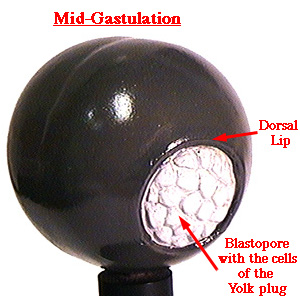
Dorsal lip of blastopore how to#
This chapter describes how to image PCP proteins in Xenopus neuroectoderm for both fixed and live samples. All of the above techniques can be applied to the analysis of the Xenopus neural plate to study the dynamics of tissue polarization, making this system one of the best vertebrate PCP models. Believed to be critical for signaling, this segregation is studied by a variety of techniques, such as direct immunostaining and imaging of fluorescent PCP protein fusions or fluorescence recovery after photobleaching (FRAP). PCP is detected by the asymmetric distribution of core PCP proteins at different borders of epithelial cells. Genetic studies in Drosophila identified several core PCP genes, whose products function together in a signaling pathway that regulates cell shape, epithelial tissue organization and remodeling during morphogenesis. Planar cell polarity (PCP) refers to coordinated cell polarization in the plane of the tissue. Last, we show that increase of SE resistive forces is detrimental for NP morphogenesis, showing that correct SE development is permissive for NTC. Its movement towards the midline is passive and driven by forces generated through NP morphogenesis. We go on to show that the SE is mechanically coupled with the NP providing resistive forces during NTC. AC occurs after CE throughout the NP and is the sole contributor of anterior NTC. CE is essential for correct positioning of the NP rostral most region in the midline of the dorsoventral axis. PCP-driven CE is restricted to the caudal part of the neural plate (NP) and takes place during the first stage. We show that NTC is a two-stage process and that CE and AC do not overlap temporally while their spatial activity is distinct. Here, using Xenopus laevis embryos, live imaging, single-cell tracking, optogenetics, and loss of function experiments we examine the contribution of convergent extension (CE) and apical constriction (AC) and we define the role of the surface ectoderm (SE) during NTC. Neural tube closure (NTC) is a fundamental process during vertebrate embryonic development and is indispensable for the formation of the central nervous system. These observations suggest that neuroectodermal PCP is not instructed by a preexisting molecular gradient, but induced by a signal from the dorsal blastopore lip. The PCP cue did not depend on the orientation of the graft and was distinct from neural inducers. Tissue transplantations indicated that PCP is triggered in the neural plate by a planar cue from the dorsal blastopore lip. By imaging Vangl2 and Prickle3, we show that PCP is progressively acquired in the neural plate and requires a signal from the posterior region of the embryo. Here we investigate the Xenopus neural plate, a tissue that has been previously shown to exhibit PCP. Although the segregation of PCP components to opposite cell borders is believed to play a critical role in this pathway, whether PCP derives from egg polarity or preexistent long-range gradient, or forms in response to a localized cue remains a challenging question. Coordinated polarization of cells in the tissue plane, known as planar cell polarity (PCP), is associated with a signaling pathway critical for the control of morphogenetic processes.


 0 kommentar(er)
